
How 3D Bioprinting Could Transform Tissue Engineering and Medical Research
Every year, thousands of people die while waiting for an organ transplant. In the United States alone, more than 100,000 people are on the waiting list for an organ, and there are far fewer donors than patients. It is one of medicine’s oldest problems: the body can fail in ways that only a replacement part can fix.
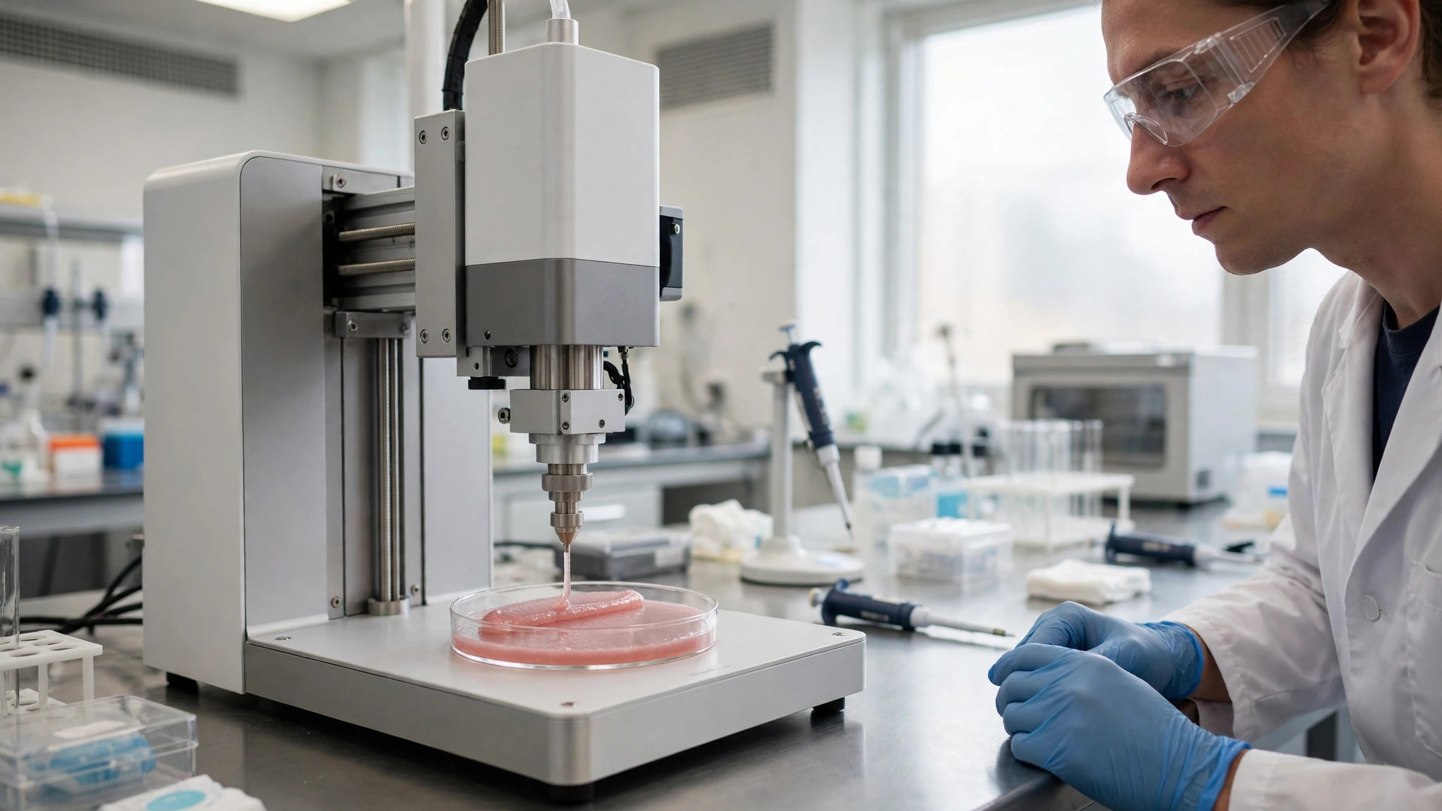
3D bioprinting offers a different answer. Instead of waiting for a donor, doctors could one day print the tissue a patient needs, using that patient’s own cells. The idea sounds like science fiction, but the machines that do it already exist, and they are working in laboratories around the world.
This article explains how 3D bioprinting works, what has already been printed, where the technology stands today, and what still has to be solved before a printed organ can save a life.
What 3D Bioprinting Actually Is
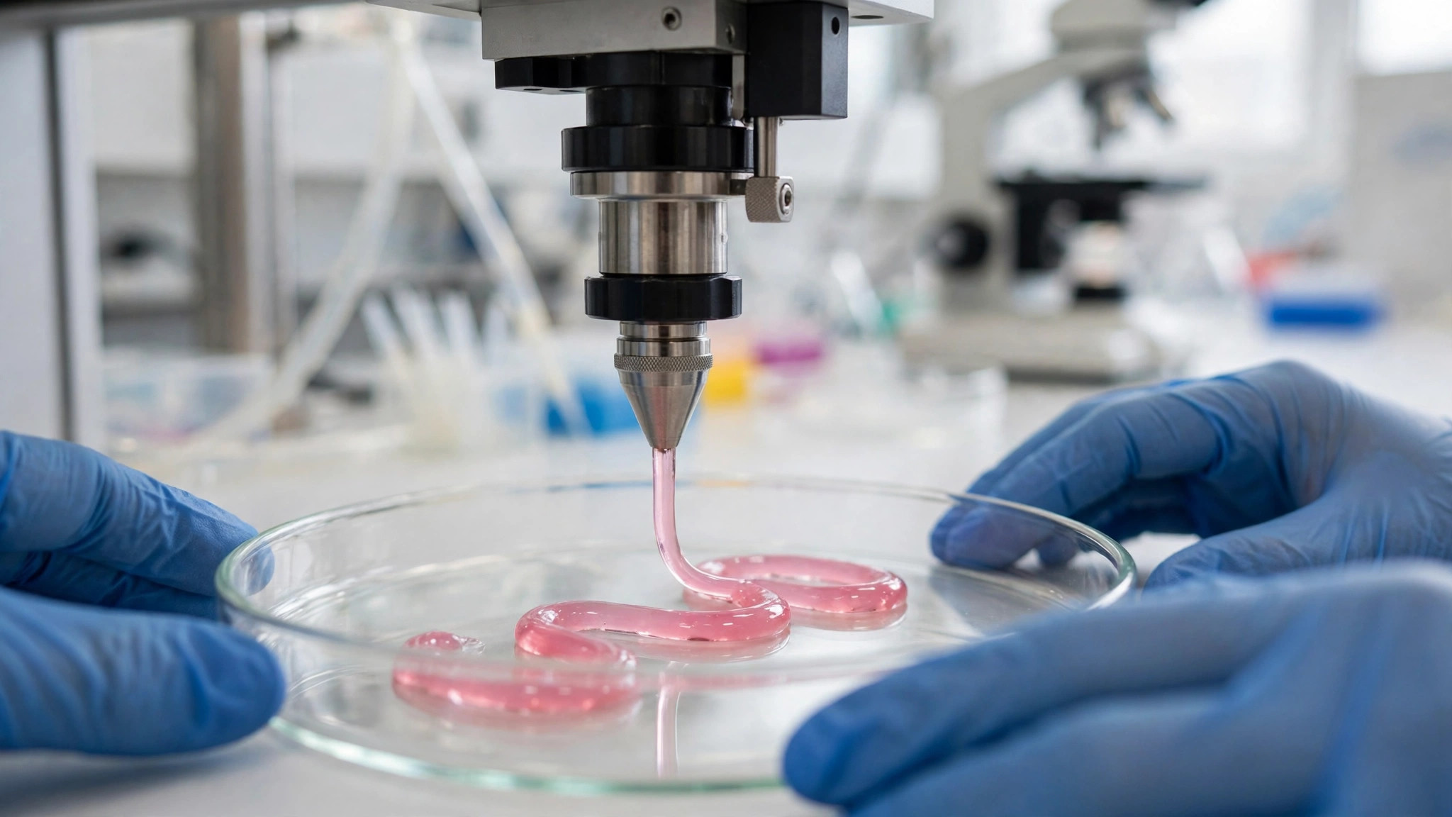
3D bioprinting is a form of 3D printing that builds living tissue instead of plastic or metal. A bioprinter lays down tiny droplets or thin lines of a material called a bioink, layer by layer, following a digital design. The bioink contains living cells, and once printed, the cells grow, connect, and begin to behave like real tissue.
The difference from ordinary 3D printing is the cargo. A normal printer works with melted plastic or powdered metal. A bioprinter works with living cells, which are fragile. They cannot be heated, crushed, or dried out, or they die. Every part of the process, from the ink to the nozzle, has to keep the cells alive.
The result is not a finished organ straight off the machine. It is a living structure that still has to mature. Printed tissue is usually placed in a bioreactor, a warm, controlled chamber that feeds it nutrients and gives it time to grow into something that works like the real thing.
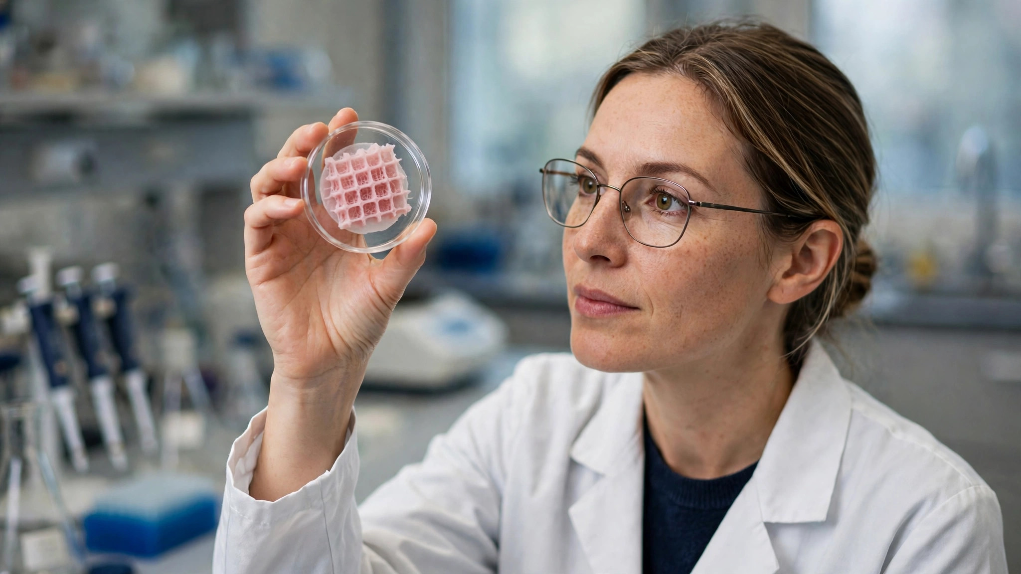
Tissue engineering is the wider field this belongs to. For decades, researchers have grown tissue in the laboratory by seeding cells onto scaffolds, which are sponge-like structures that give cells a shape to grow on. Bioprinting is a newer, more precise way of doing the same job: instead of letting cells find their own arrangement, the printer places them exactly where they belong.
How a Bioprinter Builds Living Tissue
A bioprinting project starts with a design. Researchers take a scan of the tissue they want to build, often from an MRI or CT image, and turn it into a three-dimensional model. Software then slices that model into hundreds of thin layers, each one a blueprint for a single pass of the printer.
The bioink is the heart of the process. Most bioinks are hydrogels, soft, water-rich materials that feel a little like jelly. They are made from substances the body already knows, such as collagen, gelatin, alginate from seaweed, or decellularised tissue from a donor organ with the cells removed. Living cells are mixed into the gel before printing, so every drop carries its own building crew. Researchers are also working on materials that can repair themselves after damage, known as self-healing materials, which could one day make printed tissue more durable.
There are three main ways to print. Extrusion bioprinting works like a very precise icing bag: the bioink is pushed through a fine nozzle in a continuous line. It is the most common method, because it handles thick inks and large structures well.
Inkjet bioprinting works more like an office printer, firing tiny droplets of bioink onto a surface. It is faster and gentler on cells, but it only works with thin, runny inks, which limits what it can build.
Laser-assisted bioprinting uses a pulse of light to push a droplet of bioink from a ribbon onto the surface below. It is the most precise of the three, and it can place single cells, but it is slower and more expensive.
A fourth approach, light-based printing, hardens a liquid bioink with patterned light, layer by layer. It can make very fine details, which matters for delicate structures such as blood vessels.
After printing, the tissue is not ready. It goes into a bioreactor, where it is kept warm, fed, and sometimes gently stretched or squeezed to encourage the cells to mature. A printed piece of heart tissue, for example, may be made to beat in the bioreactor before it is ever used. This maturing step can take days or weeks, and it is as important as the printing itself.
What Has Been Printed So Far
Bioprinting has already produced real results, though none of them is yet a transplantable organ. The progress so far is best understood in stages, from simple to complex.
| Tissue or structure | What has been achieved | Why it matters |
|---|---|---|
| Skin | Printed skin grafts tested in laboratories and early clinical work | Could help burn patients and wound healing |
| Cartilage | Ear-shaped cartilage printed and implanted in early trials | Cartilage has no blood vessels, so it is easier to keep alive |
| Bone | Printed bone scaffolds used in research and animal studies | Bone is rigid and can be printed with strong materials |
| Liver tissue | Small printed liver models used for drug testing | Lets researchers test medicines without animal testing |
| Heart tissue | Small beating patches of heart muscle grown in the laboratory | A step towards repairing damaged hearts |
| Kidney structures | Early printed kidney tissue and mini-kidneys for research | Kidneys are among the most needed transplants |
Skin was one of the first successes. In 2022, researchers at the Wake Forest Institute for Regenerative Medicine reported a bioprinter that could print skin directly onto a wound, using the patient’s own cells. The printers themselves are part of a wider wave of robotics innovations that are bringing automated machines into laboratories and hospitals. Skin is a good early target because it is thin, flat, and does not need a complex blood supply to survive.
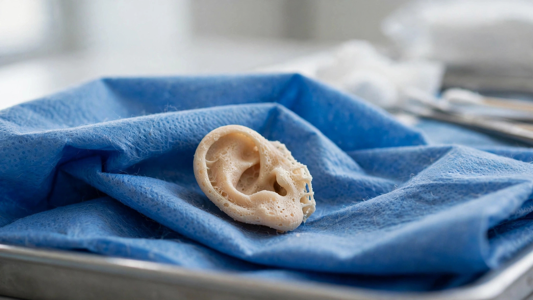
Cartilage has gone further than any other tissue towards patients. In 2022, the company 3DBio Therapeutics began a clinical trial of AuriNovo, a 3D-printed ear made from a patient’s own cartilage cells, for people born with a small or missing ear. Cartilage works well for a first implant because it has no blood vessels of its own: the printed cells can survive on nutrients from the surrounding tissue.
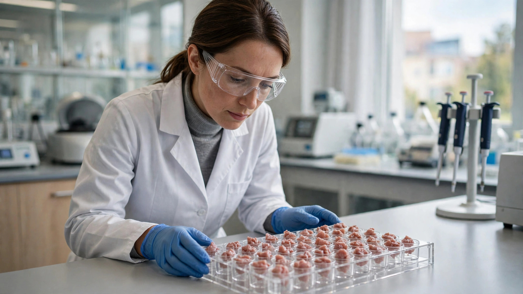
The most useful application today is not in patients at all, but in the laboratory. Printed mini-livers and other tissue models are used to test new medicines. A drug that looks promising in a petri dish can behave very differently in a real liver, and a printed tissue model gives researchers a much better test. The United States passed a law in 2022, the FDA Modernization Act 2.0, that allows drug makers to use alternatives to animal testing, which has given this work a strong push.
Where the Technology Stands Today
By 2026, bioprinting is a serious research field with real products, but it is still closer to the laboratory than the operating theatre. The machines have become cheaper and more capable, and many university hospitals now have a bioprinter of their own. All of this work depends on computing power, and the data centres that provide it are themselves changing, with modular data centers being built to meet the growing demand for AI.
The strongest area is drug testing and disease research. Pharmaceutical companies use printed tissue to test medicines earlier and more cheaply than before. Researchers also print small models of tumours to study how cancers grow and how they respond to treatment, using cells taken from a real patient. That means a treatment can be tested on a copy of a patient’s tumour before it is given to the patient.
Regenerative medicine is the next frontier. Printed skin and cartilage are in early clinical trials, and printed bone grafts are being studied for repairing injuries. These are all tissues that are relatively simple: they are thin, or stiff, or they do not need a rich blood supply.
The hardest problem, and the one that decides whether whole organs can ever be printed, is blood vessels. A living organ is full of tiny vessels that carry oxygen and nutrients to every cell. Without them, cells in the middle of a printed organ starve and die. Researchers have printed vessel-like tubes and networks of channels, but building a complete, working vascular system inside a printed organ remains unsolved.
Scale is the second problem. Printing a small patch of tissue takes minutes. Printing something the size of a human organ would take far longer, and the cells printed first would have to survive for hours while the rest is built. Keeping everything alive during a long print is a real engineering challenge.
What It Could Mean for Medicine
If those problems are solved, the change would be enormous. The most obvious benefit is an end to the organ waiting list. An organ printed from a patient’s own cells would not be rejected by their immune system, so there would be no need for a lifetime of drugs to stop rejection.
There is a nearer benefit that matters just as much. Printed tissue could make medicine personal. A doctor could test a drug on a printed model of a patient’s own tissue before prescribing it, instead of guessing from averages. For cancer patients, that could mean finding the treatment that works first time, rather than by trial and error.
It could also change how surgeons train and plan. A printed copy of a patient’s heart or liver, made from their scan, would let a surgeon practise a difficult operation before doing it for real. Some hospitals already use printed plastic models for this. A living model would be the next step.
The research benefits reach further. Scientists studying diseases such as Alzheimer’s and diabetes already use printed tissue models to watch how a disease develops in a living sample. That is something a flat layer of cells in a dish cannot show. In the longer term, researchers are even exploring whether living cells themselves could process information, the idea behind biocomputing.
None of this will arrive all at once. Printed skin and cartilage are the near term. Printed patches to repair a damaged heart or liver are the middle term. A whole printed organ is still years away, and honest researchers say so. But the direction is clear: medicine is moving from replacing broken parts with donor parts to growing the parts a patient needs.

Practical Tips / Examples / Things to Know
- If you hear about a “printed organ” in the news, check what was actually printed: a tissue model for research is very different from an organ ready for transplant.
- Cartilage and skin are the tissues closest to patients, because they survive without a complex blood supply.
- Drug testing is the biggest real-world use of bioprinting today, and it is already reducing the need for animal testing.
- A bioprinter is only half the story: the bioreactor, where printed tissue matures, matters just as much as the printing itself.
- Bioinks are made from materials the body already knows, such as collagen and gelatin, which is why printed tissue can be accepted by the body.
- If you are considering a treatment described as involving bioprinting, ask whether it is part of a registered clinical trial, and discuss it with your doctor.
Common Mistakes / Considerations / What to Avoid
- Assuming a printed organ is available today: no whole organ has been bioprinted and transplanted into a patient.
- Believing every news headline about a “breakthrough”: most bioprinting results are early laboratory work, not treatments.
- Thinking bioprinting is just normal 3D printing with different ink: living cells make the process far more delicate.
- Underestimating the blood vessel problem: without vessels, cells in the middle of a printed organ cannot survive.
- Ignoring regulation: printed tissue for patients must pass the same safety tests as any other medical treatment.
- Expecting printed organs to be cheap soon: the machines, the bioinks, and the maturing process are all expensive today.
Final Thoughts
3D bioprinting began as an idea borrowed from ordinary 3D printing, and it has grown into one of the most hopeful fields in medicine. It has already changed how new drugs are tested, it is moving printed skin and cartilage towards patients, and it offers a real path to ending the organ waiting list.
The obstacles are serious, and the blood vessel problem in particular will not be solved by a single breakthrough. But the progress so far has been steady rather than speculative: each year, the printed tissues get bigger, more complex, and closer to working like the real thing. For patients waiting for a transplant, that steady progress is the most hopeful news of all.